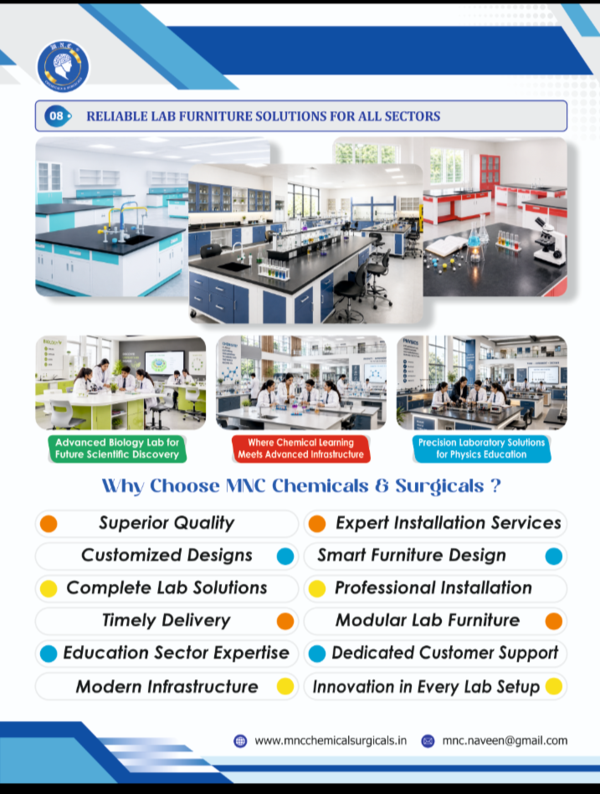
Description
The human uterus is a hollow, muscular, and pear-shaped organ within the female pelvis located between the bladder and rectum. Its primary functions include supporting menstruation, nurturing fertilized eggs, and enabling fetal development until childbirth.
Structural Features
The uterus is typically 7.5–8 cm long and about 5 cm broad, with a wall thickness of approximately 2–3 cm.
It consists of four major regions: fundus (the broad, curved upper area), body (the main, central portion), isthmus (a narrow neck region), and cervix (the lower segment projecting into the vagina).
The organ is held in position by ligaments, including the pubocervical, cardinal, and uterosacral ligaments.
Wall Layers
The uterus has three distinct wall layers:
Endometrium: The mucosal inner lining that undergoes cyclic changes, including menstruation. It comprises a functional layer that sheds during menstruation and a basal layer that remains.
Myometrium: The thick, middle muscular layer primarily responsible for contractions during labor, composed of smooth muscle arranged in three sublayers.
Perimetrium: The outer serous layer derived from peritoneum that covers most of the uterus.
Anatomical Connections
The uterus connects laterally to the fallopian tubes, through which eggs travel from the ovaries into the uterine cavity.
Inferiorly, the cervix opens into the vagina, forming part of the birth canal.
Its fundus is situated above the entry points of the fallopian tubes, while the cervical canal serves as the narrow passage between the uterine cavity and vagina.
Functional Importance
The uterus is vital for menstrual and reproductive health, serving as the site for implantation of a fertilized egg and providing a nurturing environment for fetal growth during pregnancy.
It is also involved in the labor process by contracting to facilitate childbirth.
This description is suitable for educational product catalogs and scientific documentation concerning anatomical models or illustrations of the human uterus.