
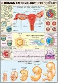
Human Embryology (Early Stage) (50x70cm)
Description
The “Human Embryology (Early Stage) (50x70cm)” chart is a mid-sized educational wall poster focused on the earliest phases of human development from fertilization through the embryonic period. This chart is ideal for classrooms, laboratories, and clinical environments, visually presenting the key morphological events that occur from conception to the eighth week.
Key Features
The chart illustrates sequential stages including fertilization, zygote formation, cleavage, morula, blastocyst development, implantation, and gastrulation.
Diagrams highlight the formation of the bilaminar and trilaminar germ discs (ectoderm, mesoderm, endoderm), neural groove, somites, and early placental structures.
Labeled illustrations may cover Carnegie stages or a week-by-week breakdown, showing visible structures like the beginning neural tube, heart tube, limb buds, and somite segmentation.
The chart frequently presents a timeline or flow of embryonic development, and often includes cross-sections or magnifications of key events, such as the formation of the amniotic cavity, yolk sac, and early chorionic villi.
Educational Use
Well-suited for high school, undergraduate, or medical instruction covering human reproductive biology or embryology.
The 50x70cm format is compact yet large enough for visible group study and reference, providing essential visual context for complex early developmental biology.
Useful for both students and educators to explain the origins and organization of basic body systems.
Specifications
| Model No. | N-C010 |
|---|


