
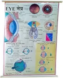
Human Eye (70x100cm)
Description
The “Human Eye (70x100cm)” chart is a large, highly detailed anatomical poster that provides comprehensive illustrations and labeling of the structure, function, and visual pathways of the human eye. It is suitable for classrooms, clinics, and visual science education, ensuring anatomical accuracy and clarity.
Key Features
The chart offers several high-quality, color-coded diagrams from different perspectives, including:
Lateral view showing lens, iris, cornea, sclera, retina, optic nerve, and extraocular muscles
Anterior (front) view of the eye and muscles
Cross-section of the eyeball highlighting all internal layers
Optic fundus (the back of the eye with blood vessels and optic disc)
The visual pathway in the brain, from retina to visual cortex
Essential anatomical structures are labeled in English (and sometimes German or Latin), with durable plastic foil and metal edging for long-term display.
Many charts also cover vision mechanics (how light passes through cornea/lens to retina), common refractive errors (myopia, hyperopia, astigmatism), and images illustrating normal vs. impaired vision.
Educational Use
Ideal for secondary, undergraduate, and clinical education, as well as for optometry and ophthalmology patient instruction.
The large 70x100cm size ensures visibility of minute anatomical details even by large groups in teaching or laboratory settings.
Laminated for protection, washable for hygiene, and often with multilingual labeling for international learning environments.
Specifications
| Model No. | N-C026 |
|---|


