
Meiosis(Animal Cell Divison) Small Size
Description
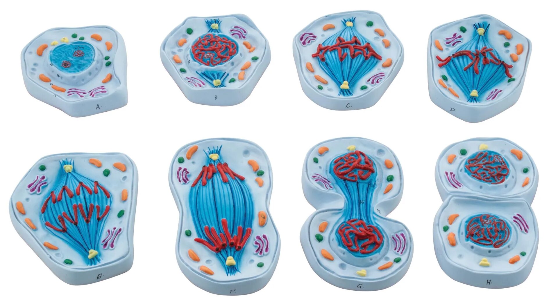
A small-size model of meiosis (animal cell division) presents a compact, visually engaging set of replica stages that highlight the critical phases of gamete formation while occupying minimal space, making it ideal for tabletop teaching or student demonstrations.
Features of Small-Size Meiosis Model
Stage Representation: Displays distinct phases of meiosis I and II—such as prophase I, metaphase I, anaphase I, telophase I, prophase II, metaphase II, anaphase II, and telophase II—often in 8–12 sequential, color-coded blocks or plaques for side-by-side comparison.
Chromosome Dynamics: Highlights chromosome pairing, crossing over (genetic recombination), segregation of homologous chromosomes, and separation of sister chromatids.
Structural Clarity: Key cellular elements—nucleus, spindle fibers, centromeres, and cell membrane—are clearly marked; models may show haploid cell outcome and differences from mitosis.
Educational Value: Designed for hands-on use in classrooms, with concise labelling, rugged build, and portability.
Meiosis Process Overview
Meiosis I reduces diploid cells to haploid by separating homologous chromosomes.
Meiosis II divides these cells again, separating sister chromatids, resulting in four genetically diverse gametes essential for sexual reproduction in animals.
Small-size meiosis models offer effective, space-efficient displays of the complex multi-phase process of animal cell division, ideal for foundational genetics education.
Specifications
| Model No. | N-EM088 |
|---|


