T.S Dicot Leaf
Description
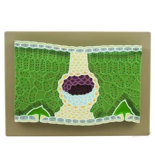
A T.S. (transverse section) of a dicot leaf—like those of bean, sunflower, or rose—reveals the classic “dorsiventral” structure, with clear differentiation between the upper and lower surfaces and distinct tissue organization.
Key Anatomical Features
Upper Epidermis: A single layer of tightly packed parenchymatous cells, typically with a thicker cuticle and fewer stomata. Provides a protective barrier and reduces water loss.
Lower Epidermis: Single cell layer, thinner cuticle, with abundant stomata for gas exchange. Stomata are kidney-shaped and more numerous in the lower epidermis, making the leaf hypostomatic.
Mesophyll: Divided into two regions:
Palisade Parenchyma: Just beneath the upper epidermis, made of elongated and closely packed cells, rich in chloroplasts—main site for photosynthesis.
Spongy Parenchyma: Below the palisade, consisting of loosely packed, irregular cells with large intercellular spaces for gas exchange.
Vascular Bundles: Present as veins and midrib, containing xylem (towards the upper epidermis) and phloem (towards the lower epidermis), surrounded by a bundle sheath of parenchymatous cells.
Venation: Reticulate (net-like), characteristic of dicot leaves.
Specifications
| Model No. | N-EM0129 |
|---|


